
What a Child’s Pupils Can—and Cannot—Tell You After a Head Injury
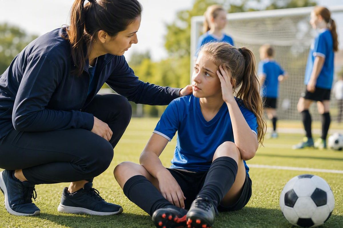
Equal dilation does not establish concussion, and normal-looking pupils do not rule it out. A newly unequal pupil after a head injury needs emergency care.
The immediate answer: when pupil changes require emergency care
Act now: If one pupil becomes newly larger than the other after a head injury, call 911 or your local emergency number, or go to an emergency department. Do not keep the child at home while repeatedly checking the pupil, comparing photographs, measuring the eyes, or trying a phone application.
A newly unequal pupil after a hit, fall, collision, or other injury that rapidly moved the head is an emergency warning sign. It may indicate an injury more serious than an uncomplicated concussion, although the pupil change does not identify the injury by itself. Mass General Brigham advises emergency care when one pupil becomes larger than the other after a child hits their head, especially when other neurological warning signs are present (see its pediatric concussion guidance).
Pupils that appear newly and unusually enlarged after head trauma also require emergency assessment, particularly if the child is deteriorating or the pupils appear not to change normally as conditions change. Do not assume the situation is safe simply because both pupils look the same size. Mayo Clinic lists pupils that are larger than usual or unequal among the reasons to seek emergency care after a head injury (review its emergency guidance).
Seek emergency help immediately if a pupil abnormality occurs with any of these danger signs:
- a headache that keeps worsening or does not go away;
- repeated vomiting;
- a seizure or convulsions;
- weakness or numbness;
- slurred speech;
- poor balance or coordination;
- increasing confusion;
- major or unusual behavior changes;
- prolonged loss of consciousness; or
- difficulty waking or staying awake.
These are danger signs, not a checklist for diagnosing concussion. Abnormal pupils can occur with conditions more serious than concussion, and emergency clinicians must determine the cause.
Do not delay care to find an old photograph, compare the eyes under different lamps, measure the pupils, shine a flashlight into them repeatedly, or download an eye-testing application.
Online information cannot determine the severity of an individual child’s head injury. If the child is getting worse, cannot be awakened normally, or has a new pupil abnormality, contact emergency services rather than continuing an at-home assessment.
Do dilated pupils mean someone has a concussion?
No. Dilated pupils alone neither confirm nor rule out a concussion.
Visible dilation—especially when both pupils are equally enlarged—is nonspecific. Pupils change size naturally, and their appearance can be affected by lighting, emotional arousal, medications, substances, eye conditions, and other factors. Michigan Medicine specifically identifies the belief that dilated pupils reliably indicate concussion as a misconception and emphasizes that concussion symptoms vary from person to person (read its clinician-led myth review).
Some secondary health-information sources include dilation among possible findings following concussion. That means dilation may be observed in some injured people; it does not establish that dilation is typical, unique to concussion, or accurate enough for self-diagnosis.
The reverse is equally important: normal-looking pupils do not exclude concussion. A child may have equal, apparently responsive pupils while experiencing headache, dizziness, confusion, light sensitivity, nausea, balance difficulty, or concentration problems.
Concussion primarily affects brain function, so many effects are experienced by the injured person rather than seen by an observer. A coach may notice that a player looks dazed or answers slowly, but a child can also look outwardly normal while describing head pressure, blurred vision, mental fog, or unusual fatigue.
Pupil observation is therefore useful mainly for triage:
- A newly unequal or conspicuously abnormal pupil can signal an emergency.
- Equal dilation does not establish concussion.
- Normal-looking pupils do not provide medical clearance.
- A pupil check cannot determine whether an athlete may return to play.
The practical question is not, “Do these pupils prove concussion?” It is, “Is there a new pupil abnormality or another danger sign that makes this an emergency—and, if not, does the injury still require professional assessment?”
Equal dilation, unequal pupils, and poor light response are different findings
Pupil size, symmetry, and responsiveness are related but distinct observations:
- Size describes how large each pupil appears.
- Symmetry describes whether the pupils are approximately the same size.
- Responsiveness describes how their size changes under different visual and lighting conditions.
Two large but equal pupils are not the same finding as one pupil suddenly becoming much larger. Likewise, absolute size does not establish whether a pupil’s response is normal.
| What you notice | What it may mean | Appropriate response after a head injury |
|---|---|---|
| Both pupils appear equally enlarged | Dim surroundings, stress, excitement, nervousness, anxiety, medications, substances, and other factors can affect both pupils. Equal dilation is not specific to concussion. | Do not treat equality as proof that everything is fine. Newly unusual enlargement after trauma requires emergency assessment, particularly if the child is worsening or the pupils appear poorly responsive. |
| One pupil is newly larger than the other | This is anisocoria. Possible causes include eye, nerve, or brain problems. | Call emergency services or go to an emergency department. Do not delay for repeated home testing. |
| A pupil appears poorly responsive as conditions change | Responsiveness is different from size. An apparently abnormal response may have neurological, eye-related, medication-related, or testing-related explanations. | After trauma, obtain immediate professional assessment rather than trying to interpret repeated flashlight tests at home. |
| There has always been mild asymmetry | Some people have a small physiological difference between their pupils. | Tell the clinician if the difference is known to be longstanding. Do not rely on that explanation if it appears new, has changed, or accompanies post-injury symptoms. |
| The pupils look normal | Normal appearance does not rule out concussion or every serious injury. | Consider the injury and the full symptom pattern. Remove a possibly concussed athlete from play and arrange professional evaluation. |
Anisocoria is the medical term for unequal pupil size. It describes an observation, not a diagnosis. Head or eye trauma can cause anisocoria, but so can several unrelated conditions. The underlying cause determines what testing or treatment is appropriate, and sudden anisocoria with concerning symptoms needs prompt evaluation (see Cleveland Clinic’s overview).
Both pupils normally enlarge in dim light. Sympathetic arousal associated with stress, excitement, nervousness, or anxiety can also make them appear large. These alternatives help explain why equal dilation is a poor standalone concussion sign, but they should not be used to dismiss a new change after a collision or fall.
A small, longstanding difference may be physiological. The difficulty is that a caregiver usually cannot determine safely at home whether newly noticed asymmetry is benign, eye-related, medication-related, or neurological. After trauma, respond to new anisocoria as an emergency rather than trying to identify its cause yourself.
Avoid relying on a millimeter cutoff. A photograph or casual ruler measurement cannot establish the cause or severity of a head injury.
Similarly, do not repeatedly shine a light into the child’s eyes. An informal flashlight check is not equivalent to a controlled neurological examination, and a response that seems normal to a caregiver cannot clear the child.
Symptoms that matter even when the pupils look normal
Concussion symptoms can be obvious or subtle and can affect the body, thinking, mood, sleep, and vision. The following are possible symptoms, not proof that a concussion occurred.
Physical symptoms
- headache or pressure in the head;
- dizziness;
- nausea or vomiting;
- balance difficulty or unsteadiness;
- fatigue or low energy;
- sensitivity to light or noise; and
- feeling slowed down or generally unwell.
Thinking and memory symptoms
- confusion or feeling “foggy”;
- difficulty concentrating;
- memory gaps around the event;
- slow answers or difficulty following instructions;
- repeated questions; and
- difficulty completing familiar schoolwork or tasks.
Emotional or behavioral symptoms
- irritability;
- unusual sadness or nervousness;
- emotional reactions that seem out of character;
- withdrawal from usual activities; and
- behavior that differs noticeably from the child’s baseline.
Sleep-related symptoms
- sleeping more or less than usual;
- difficulty falling asleep;
- unusual drowsiness; and
- changes in the normal sleep pattern.
Visual symptoms
- blurred or double vision;
- light sensitivity;
- difficulty focusing;
- eye strain;
- trouble following a moving object; and
- difficulty reading or using screens.
Loss of consciousness is not required for concussion, and most concussions do not involve it. Concussion can also follow an impact to the body or rapid acceleration and deceleration of the head without a direct blow to the skull. Symptoms may begin immediately or become noticeable over the following hours or days (Mayo Clinic summarizes these mechanisms and symptom patterns).
A child may initially be distracted by the game, frightened by the fall, or unable to describe what feels wrong. An initially normal appearance is therefore not definitive.
Do not confuse common possible concussion symptoms with emergency danger signs. A mild headache or brief dizziness may prompt removal from play and professional evaluation. A worsening persistent headache, repeated vomiting, seizure, weakness, slurred speech, increasing confusion, marked coordination problems, or difficulty waking requires emergency care.
Neither “They look fine” nor “Their pupils look normal” is enough to clear a child after a concerning impact.
What to do after a suspected sports concussion
If an athlete may have a concussion but has no emergency warning signs, take the following steps.
1. Remove the athlete from play immediately.
Stop the game, drill, practice, bike ride, or other activity that could produce another head impact. Do not ask the child to try another play to see how they feel. Removal is a safety response, not a diagnosis. Pediatric concussion guidance advises removing a child from risky activity and obtaining prompt medical care rather than relying on sideline appearance (see Mass General Brigham’s guidance).
2. Do not use a pupil check as a return-to-play test.
Normal pupils do not mean the brain is uninjured.
A player should not return to an activity carrying another head-injury risk based on a coach’s observation, a parent’s flashlight check, a phone application, a teammate’s opinion, or the child’s desire to continue.
3. Arrange a prompt healthcare evaluation.
For a child with a possible concussion, the CDC advises assessment by a healthcare provider within the first 24 to 48 hours. The provider can review the injury, symptoms, observed changes, and medical history and determine what examination or follow-up is appropriate (follow the CDC’s pediatric guidance).
That pediatric timeline should not be applied mechanically to adults or older adults. Age, medications, medical history, injury mechanism, and symptom severity can affect urgency. Emergency warning signs always override routine appointment timing.
4. Monitor for changes.
Pay attention to how the child acts and feels because symptoms can emerge or become recognizable later. Useful information for the clinician includes:
- when and how the injury occurred;
- whether there was a direct head impact or rapid body movement;
- symptoms noticed immediately and later;
- whether symptoms are improving or worsening;
- any loss of consciousness or memory gap;
- known baseline pupil asymmetry;
- relevant medications or substances; and
- observations from coaches, officials, teammates, or witnesses.
Writing down this information may help, but documentation must never delay emergency care.
5. Escalate if a danger sign appears.
A newly unequal or unusually enlarged pupil, worsening headache, repeated vomiting, seizure, weakness, numbness, slurred speech, poor coordination, increasing confusion, major behavior change, prolonged loss of consciousness, or difficulty waking changes the response. Stop routine monitoring and obtain emergency help.
6. Follow an individualized return plan.
Sleep, school, screen use, exercise, and return-to-sport decisions depend on the child’s symptoms and clinical assessment. There is no single home protocol appropriate for every child.
Clearance for contact practice, competition, or another activity carrying head-injury risk should follow professional assessment—not normal-looking pupils or a home symptom check.
Eye and vision problems after concussion
Pupil size is only one possible eye-related observation. Concussion can affect how a person sees, focuses, and coordinates the eyes even when the eyes look normal from the outside.
Possible symptoms and findings include:
- blurred or double vision;
- sensitivity to light;
- difficulty focusing at near or far distances;
- trouble tracking a moving person, ball, or line of text;
- atypical eye movements;
- eye misalignment;
- eye strain; and
- possible eyelid drooping.
Many of these are experienced symptoms rather than changes a parent or coach can see. A child may say that words move on the page, bright light hurts, the classroom feels visually overwhelming, or switching focus between a notebook and the board is difficult.
One possible problem is convergence insufficiency. This means the eyes have difficulty turning toward and working together on the same nearby target. It is a possible post-concussion problem, not a feature of every concussion.
Vision difficulties may become more apparent during:
- reading or homework;
- scrolling or typing on a screen;
- moving through a crowded hallway;
- watching rapid action;
- riding in a vehicle; or
- spending time in bright or visually busy surroundings.
A medically reviewed overview lists double vision, light sensitivity, tracking difficulty, atypical eye movements, eye misalignment, and drooping eyelids among possible concussion-related eye problems while emphasizing that eye appearance cannot diagnose concussion at home (review the eye-symptom overview).
Persistent double vision, light sensitivity, focusing difficulty, tracking problems, or trouble reading and using screens merits clinical follow-up. Depending on the symptoms, evaluation may involve the clinician managing the concussion and an appropriate eye-care or rehabilitation professional.
Do not assume every reading or screen problem is purely an eye issue. Headache, fatigue, attention difficulty, motion sensitivity, and visual coordination problems can overlap.
Externally visible abnormalities should be distinguished from ordinary screen intolerance. New eyelid drooping or obvious eye misalignment after trauma also needs prompt professional assessment; if either occurs with an abnormal pupil or neurological danger signs, use emergency care.
How clinicians evaluate concussion and serious brain injury
Concussion assessment begins with the event and the person—not with a single pupil observation.
A clinician may consider:
- the mechanism and timing of the injury;
- whether the head was struck or rapidly accelerated;
- symptoms reported by the injured person;
- changes observed by others;
- loss of consciousness or memory;
- previous concussions;
- neurological, eye, migraine, and other medical history;
- medications and other possible contributors; and
- whether symptoms are improving, stable, or worsening.
The physical and neurological examination may include mental status, speech, memory, balance, coordination, strength, sensation, vision, eye movements, and pupil findings. These observations are interpreted together. One normal element does not necessarily cancel out abnormalities elsewhere.
There is no conclusive at-home eye test for concussion. Casual inspection cannot reproduce a clinical examination, and a child’s ability to follow a finger, identify a number, or read text does not establish clearance.
Imaging has a different purpose. A CT scan may be used when bleeding, skull injury, or another structural problem is suspected; it is not a simple test that confirms an uncomplicated concussion. Most children with concussion do not require CT or MRI, but this is not a universal rule, and the decision depends on the individual injury and risk factors (the CDC addresses imaging in its pediatric guidance).
Normal imaging does not by itself exclude concussion. Concussion is diagnosed clinically from the injury history, symptoms, observed changes, and examination, so a scan can be normal while the person has genuine concussion symptoms.
Not every person with unequal pupils will undergo the same tests. Depending on the suspected cause, evaluation may focus on the brain, nerves, eyes, medications, or another condition.
Pupil appearance is therefore one piece of triage information. It may identify a situation requiring emergency attention, but it cannot name the injury, determine its severity, or replace a complete examination.
What pupil-reflex research can—and cannot—show
A parent looking at a child’s eyes is making a casual visual observation. Quantitative pupillometry is different: it uses controlled equipment and testing conditions to measure features of the pupillary light reflex.
Depending on the system, measurements may include:
- baseline or maximum pupil diameter;
- minimum diameter after a light stimulus;
- latency before constriction begins;
- constriction velocity;
- dilation velocity; and
- recovery time.
These are dynamic measurements collected over time. They are not equivalent to asking whether the pupils look large.
Research has found associations between concussion history and several instrument-measured pupil-reflex features. One retrospective clinical review used an iPhone-based measurement system to compare latency, pupil diameters, constriction velocity, and recovery time in people with and without concussion histories. Differences appeared in several measures, but the study design showed associations; it did not establish that an application could diagnose an individual concussion (read the peer-reviewed study).
A scientific review of mild traumatic brain injury described measured pupil responses as delayed, slowed, reduced, and generally symmetrical. Its summary reported smaller—not larger—baseline pupil diameter in mild TBI. That evidence does not support treating obvious bilateral dilation as a characteristic standalone sign of uncomplicated concussion (see the scientific review).
Pupil measurements may be affected by:
- lighting and adaptation to light;
- age;
- attention;
- fatigue and sleep;
- emotional state;
- focusing on a nearby object;
- medications or other substances;
- iris and other eye characteristics;
- disease; and
- equipment and testing conditions.
These influences make standardization and professional interpretation important. They also explain why a subtle change detected by a controlled instrument cannot be reduced to “the pupil looks dilated.”
Quantitative pupillometry may eventually become a useful part of a broader assessment. The available evidence supports describing it as a promising possible aid, not an established standalone diagnostic test.
In particular, retrospective research involving an application does not validate consumer phone testing for:
- deciding whether to call emergency services;
- diagnosing concussion at home;
- excluding serious brain injury;
- deciding whether imaging is needed; or
- clearing an athlete to return to play.
A phone camera may create an impression of precision without answering the important clinical questions. If one pupil becomes newly larger after an injury, emergency care should not wait for an app. If the pupils look normal but concussion symptoms are present, an app should not override those symptoms or replace professional assessment.
Frequently asked questions
Can both pupils be dilated after a concussion?
Both pupils may appear dilated after an injury, but equal dilation does not establish that a concussion occurred. Dim lighting, stress, excitement, anxiety, medications, substances, and other factors can enlarge both pupils.
If both pupils look newly and unusually enlarged after head trauma, obtain emergency assessment—particularly if they appear poorly responsive or the child has worsening neurological symptoms. Do not dismiss a new post-injury change simply because both pupils look the same.
Is one pupil larger than the other after a head injury an emergency?
Yes. If one pupil becomes newly larger than the other following a head injury, call emergency services or go to an emergency department.
A mild difference can be longstanding in some people, but a caregiver usually cannot determine the cause of new asymmetry safely at home. Do not postpone care while searching through photographs or measuring the pupils.
Can someone have a concussion if their pupils look normal?
Yes. Normal-looking pupils do not rule out concussion. Headache, dizziness, nausea, confusion, balance difficulty, memory or concentration problems, fatigue, light or noise sensitivity, mood changes, altered sleep, and blurred or double vision can occur while the pupils appear normal.
Medical clearance should never be based on pupil appearance alone.
Can concussion symptoms appear hours or days after an injury?
Yes. Symptoms can be immediate, subtle, or delayed for hours or days. A child may also recognize or report them only after returning to school, reading, using a screen, or becoming physically active.
Monitor for changes in how the child acts or feels and arrange prompt evaluation when concussion is possible. Seek emergency care immediately if a danger sign appears.
Can a phone-based pupil test diagnose a concussion?
No consumer phone-based pupil test has been established by the available evidence as a standalone method for diagnosing concussion, excluding serious injury, handling emergency triage, or granting return-to-play clearance.
Researchers can use specialized systems to measure subtle aspects of the pupil reflex, but those measurements depend on controlled conditions and professional interpretation. They are not equivalent to visually checking whether a pupil looks large.
The decision-focused takeaway is simple: Pupil appearance cannot diagnose concussion, and normal pupils do not provide clearance. A newly enlarged or unequal pupil after a head injury requires emergency care. If emergency signs are absent but concussion is possible, stop the activity, monitor for delayed changes, and arrange prompt professional assessment instead of relying on an at-home eye check.